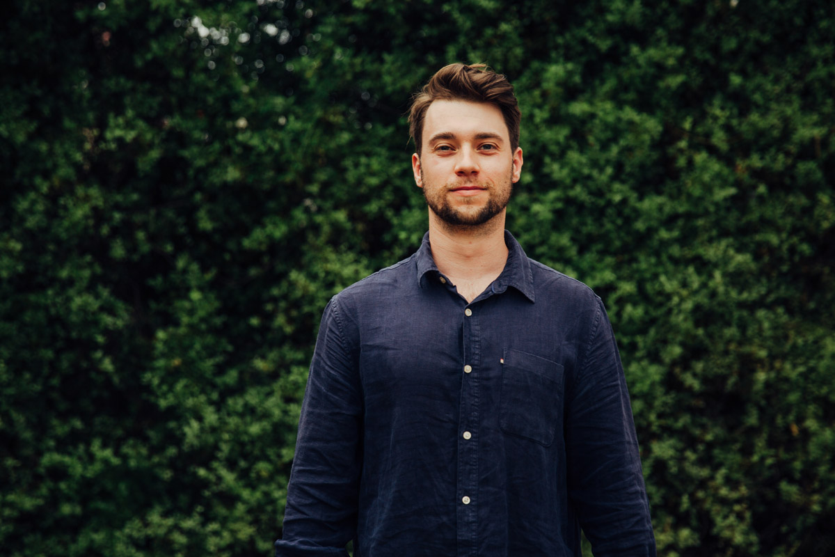
Growing ‘mini-brains’ in the lab has provided neuroscientists with unprecedented insights to better understand neurological disorders – something that PhD candidate Michael Zabolocki is taking full advantage of in his research.
What is your research is focussed on?
I’ve been fortunate enough to join Dr Cedric Bardy’s lab as a PhD candidate, where we are focussed on understanding the underlying molecular mechanisms and genetic predispositions of neurological disorders such as Parkinson’s disease, Sanfilippo syndrome and Glioblastoma, a brain cancer.
We utilise new cell-reprogramming technologies to generate human neuronal cultures from induced pluripotent stem cells (iPSCs), which are derived from human skin-cell biopsies. This gives us the ability to effectively grow a live ‘mini-brain’ within the lab from healthy patients and those with neurological disorders. Access to live human brain biopsies is limited, so our ability to generate live patient-derived neurons in-vitro has provided unprecedented insights into the mechanisms underlying neurological disorders.
These neuronal cultures are an important tool in neuroscience research, but there is room to improve their neural circuitry relative to the developed human brain. My research is focussed on exploring the electrophysiology of such human neuronal models and using innovative methods to accelerate their functional maturity. My first year was also focussed on developing the biotech product BrainPhys Imaging, which improves functional live-imaging assays on such neuronal models. I hope my research will reduce the gap between human neurons in-vitro and the human brain itself, leading to improvements in drug translation from bench to clinic.
What inspired you to pursue a PhD in neuroscience?
If you consider the neural circuitry of the human brain as a jigsaw, where every piece is a single neuron, our developed brains would comprise billions of such pieces. These fire and integrate sensory inputs in an incredibly complex manner, which gives rise to human cognition. Changes to the cellular mechanisms of these neurons can alter the pieces of this ‘puzzle’ and distort the neural circuitry of the human brain, leading to potentially devasting down-stream effects.
Unfortunately, I’ve witnessed the impact of this first-hand, as my uncle suffers from Multiple Sclerosis (MS). Watching him live with MS has sparked my fascination in exploring the molecular mechanisms underlying neurological disorders. By pursuing a PhD in neuroscience, I’m fortunate enough to have the resources and tools to do this, surrounded by researchers who are leaders within their respective fields.
Can you describe a challenge in your life and how you dealt with it?
Going through the scrutiny of the peer-review process in academic journals is difficult. Being new to the academic stream, I found this an incredibly steep learning curve and it evoked a lot of self-doubt and fear – but I tried to see a bigger picture, recognising that my work will potentially have a beneficial impact that trumps the emotions I was feeling at the time. Fortunately, my excellent supervisor/mentor, colleagues and family helped guide me through the process. I really owe a lot to the team.
What is something you are most proud of and excited for in the future?
I recently had a paper published in Nature Communications on the biotech product BrainPhys Imaging, which is now available globally via STEMCELL Technologies (DOI: 10.1038/s41467-020-19275-x). It serves as a new and improved culture medium for functional imaging assays on neuronal cultures, and is based on the formulation of BrainPhys. Currently, a lot of competitive imaging media fail to maintain the physiological activity of neurons in-vitro – a particular issue when they are used for functional imaging assays, such as calcium imaging and optogenetics, which require brain cells to fire optimally. We show that BrainPhys Imaging reduces phototoxicity and improves imaging quality across the visible light spectrum. I’m excited to see what research BrainPhys Imaging may drive for other neuroscientists.
While I’m proud of my first publication, I’m now looking forward to future collaborations and publications. As I grow as a PhD candidate, I’m learning new skills so I can dive deeper into understanding the neural circuitry of our human neuronal models. As the tools within neuroscience continue to develop, we are constantly postulating new ideas surrounding our neuronal models that unravel the cellular mechanisms underlying neurological disorders.
What does a normal day look like for you?
It’s not like in TV or movies, where data is generated almost instantaneously and research breakthroughs are produced during sudden “Eureka!” moments. My normal day comprises meetings, brainstorming with the team at Bardy Lab, data analysis, presenting information and electrophysiology-based lab work. I’m constantly being forced to think of problems critically and solve science-based issues, which involves an element of creativity. I often leave at the end of a day feeling great accomplishment in what I’ve done and what we are working towards as a team.
How do you like to relax or spend your spare time?
I spend my spare time playing and producing music. I’m fortunate enough to have a lot of talented friends I can create with. I also like to box, mountain bike ride and play soccer. The social and creative aspect of these activities provides balance in my life, which is vital to a productive and enjoyable PhD candidature.

