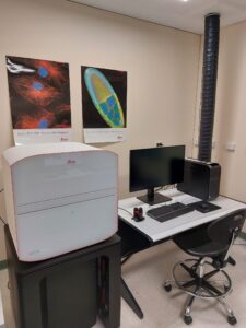
We are delighted to announce the addition of the Leica Mica confocal microscope to the light microscopy capabilities at Flinders Microscopy and Microanalysis. Supported by the expertise of Dr Nick Eyre and Pat Vilimas, researchers at the facility will be able to use the Mica for the imaging of both live and fixed samples at high resolution and high speed.

This instrument is capable of seamlessly transitioning between widefield and confocal imaging at the touch of a button. It can scan entire samples and generate rapid widefield overviews, zoom in on the region of interest, and switch to confocal imaging without requiring sample movement.
Moreover, the Mica’s intelligent automation streamlines the setup process, enabling users to initiate experiments within minutes of placing samples into the enclosed system.

The Mica’s patented spectral unmixing technology, FluoSync, and clever light path design allows simultaneous imaging of up to four different fluorophores, whether using widefield or confocal imaging. Moreover, this technology ensures that samples are maintained under ideal physiological conditions, facilitating long-term live cell imaging as needed.
Contact cmph.microscopy@flinders.edu.au to get in touch about using this instrument.