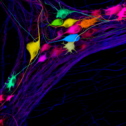
‘Networking Event’ by Ms Bao Nan Chen
First prize
This image shows nerve cells in the wall of the human colon that have been labelled using a mixture of antibodies. The immunohistochemical staining and confocal microscopy imaging were done by Ms Bao Nan Chen. The resulting image was extensively processed by Dr Tim Hibberd to create the different colours of the neurons. The work was carried out in the laboratories of Prof Simon Brookes as part of a project in the discipline of Human Physiology which was funded by the National Institutes of Health (NIH) of the USA (Stimulating Peripheral Activity to Relieve Conditions (SPARC) Program, Award Number OT2-OD024899).
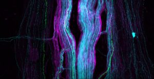
‘That’s my gut’ by Dr Michaela Johnson
Second prize
An “ascending nerve” running through the wall of the human colon. This image shows the nerve fibres of the ascending nerve (cyan) surrounded by a protective sheath (magenta) and a few myelinated nerve fibres (green).
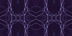
‘Painful Webs of the Womb” by Dr Kelsi Dodds
Equal third prize
This artwork is a tiled mosaic of pain-sensing nerves surrounding a blood vessel in the mouse uterus. The neural “web” created demonstrates the highly interconnected pathways by which pain may arise from the womb.
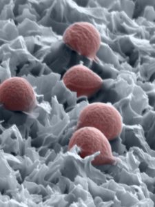
‘Arrested Development’ by Mr Richard Bright
Equal third prize
Inspired by the Dragonfly wing, hydrothermally etched biomaterials promise to be an effective first line of defence against ever increasing implant-associated infections and antibiotic resistance. The bacteria in the image seemed to be trapped and their cell walls partially warped by mechanically induced stress, eventually leading to their death. Surfaces such as the one pictured will inevitably become a staple for implanted medical devices.
The SEM image is Staphylococcus aureus on the nanospikes and was acquired at 75K mag.
To read more about SALHN Discovery Science Artwork competition click here.